UNIVERSITY OF VIRGINIA
  • Home
  • Previous Research
    • Centrifugal Microdevices >
      • PET Chips
    • Clinical Diagnostics and Forensic Analysis >
      • Cell Sorting and Solid Phase Extraction >
        • Acoustic Differential Extraction
        • Isolation of Circulating Tumor Cells
        • Enhanced Sperm Cell Recovery
        • Monolith Stationary Phase for Microfluidic DNA Purification
        • Nucleic Acid Purification in a Two-Stage, Dual-Phase Microchip
        • Large Volume Reduction Solid Phase Extraction
        • Plastic SPE Microdevices
      • Polymerase Chain Reaction (PCR) >
        • Infrared PCR
        • Microwave PCR
      • Label-Free Optical Methods for DNA and Cell Quantification
      • DNA Extraction and PCR Amplification
    • Fluidic Control >
      • Passive Valving
    • Genetic Analysis >
      • A Fully Integrated Microfluidic Genetic Analysis Device for the Detection of Blood Cancers
      • Electric Field-Flow Fractionation for DNA Concentration
      • Acousto-Optic Tunable Fiber
    • Narcotics and Explosives Colorimetric Detection
  • People
  • Publications
  • Collaborators
  • Contact
  • Conferences
Picture

A Fully Integrated Microfluidic Genetic Analysis Device for the Detection of Blood Cancers

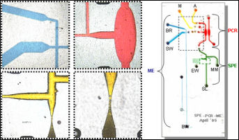
We are developing a fully-integrated microdevice capable of DNA extraction, PCR amplification and the subsequent electrophoretic separation specifically for the detection of T-cell lymphoma (TCL).  While we have recently reported a fully-integrated device for the detection of bacteria [1], the interrogation of human genomic DNA from whole blood provides new challenges, especially when detecting gene rearrangements correlative with cancer.  Detection of TCL involves the PCR amplification of select sequences in the T-cell receptor gene that are likely to have undergone gene rearrangement.  These PCR fragments represent a polyclonal cell population in normal individuals and a monoclonal cell population in patients with lymphoma in a way that can be discriminated by electrophoretic separation. In clinical labs, the PCR that follows DNA extraction requires ~3 hours followed by a 40 min capillary electrophoresis separation under single-stranded conditions and utilizing 4-color detection.  While integration of processing steps is a large advantage over traditional methods, the reduction in the times associated with these lengthy processes is also an important benefit. 

1. Easley, C.J.,  Karlinsey, J.M.,  Bienvenue, J.M., Legendre, L.A., Roper, M.G., Feldman, S.H., Hughes, M.A., Merkel, T.J., Ferrance, J.P., Landers, J.P. PNAS, 2006, 109(51), 19272-19277. 

Picture
Contact Landers Research Group
jpl5e@virginia.edu
(434) 243-8658
375, 379, 395 Chemistry Building
McCormick Road
​Charlottesville, Va 22904
UVA Chemistry Department
Powered by Create your own unique website with customizable templates.
  • Home
  • Previous Research
    • Centrifugal Microdevices >
      • PET Chips
    • Clinical Diagnostics and Forensic Analysis >
      • Cell Sorting and Solid Phase Extraction >
        • Acoustic Differential Extraction
        • Isolation of Circulating Tumor Cells
        • Enhanced Sperm Cell Recovery
        • Monolith Stationary Phase for Microfluidic DNA Purification
        • Nucleic Acid Purification in a Two-Stage, Dual-Phase Microchip
        • Large Volume Reduction Solid Phase Extraction
        • Plastic SPE Microdevices
      • Polymerase Chain Reaction (PCR) >
        • Infrared PCR
        • Microwave PCR
      • Label-Free Optical Methods for DNA and Cell Quantification
      • DNA Extraction and PCR Amplification
    • Fluidic Control >
      • Passive Valving
    • Genetic Analysis >
      • A Fully Integrated Microfluidic Genetic Analysis Device for the Detection of Blood Cancers
      • Electric Field-Flow Fractionation for DNA Concentration
      • Acousto-Optic Tunable Fiber
    • Narcotics and Explosives Colorimetric Detection
  • People
  • Publications
  • Collaborators
  • Contact
  • Conferences